SCELSE Facilities
Advanced Biofilm Imaging Facility
Unlock cutting-edge imaging potential with SCELSE’s Advanced Biofilm Imaging Facility (ABIF), where world-class technology meets interdisciplinary research excellence.
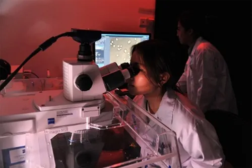
Microscopy is an indispensable tool for SCELSE’s multidisciplinary research into biofilms and microbiomes. At the Advanced Biofilm Imaging Facility (ABIF) at SCELSE, NTU, our suite of advanced microscopes also support cutting-edge imaging beyond the microbial world—including non-microbial life sciences and material sciences.
ABIF is also open to external users —researchers, innovators, and businesses alike. Gain access to state-of-the-art equipment and tap into our expertise for tailored imaging solutions. As part of the prestigious NTU Optical Bio-Imaging Centre (NOBIC) and SingaScope networks, ABIF offers seamless access to Singapore’s premier microscopy infrastructure.
Dive into the captivating results from our 2018 Imaging Contest and NOBIC Image Contest to witness the breathtaking detail and groundbreaking potential ABIF our facility can unlock. These incredible images are a testament to the precision and innovation that our cutting-edge microscopy enables.
Explore our comprehensive services and competitive pricing. Contact us today for your imaging challenges.
Email: abif@e.ntu.edu.sg; nobic.facilities@e.ntu.edu.sg
Or contact:
FOO Yong Hwee (Dr) – ABIF manager: user registration and training, consultation on sample preparation and imaging, super-resolution imaging, image processing and analysis (FIJI), etc.
- Radek MACHÁN (Dr) – NOBIC facilities manager: user registration and training, technical issues with microscopes, OMERO, FIJI, consultation on imaging, image processing & analysis, etc.
- Peter TÖRÖK (Prof) – SCELSE director of imaging, NOBIC director
| About SingaScope As a key contributor to Singapore’s premier microscopy landscape, SCELSE’s ABIF is part of the SingaScope network. This cutting-edge initiative, SingaScope, unites microscopy platforms across five leading institutes, providing scientists, including those at SCELSE, with unparalleled access to advanced imaging resources. |

Utilising State-Of-The-Art Technology
Types of Microscopes
Our facility is equipped with the following microscopes:
- Custom Raman/Brillouin microscope – a custom inverted microscope for micro-spectroscopy of inelastic light scattering
- Custom 2-photon microscope – an upright 2-photon microscope for 2-photon fluorescence and second harmonic generation imaging
- Carl Zeiss ELYRA PS.1 with LSM 780 confocal – super-resolution (SIM, PALM, STORM, …) and TIRF microscope combined with a laser scanning confocal
- Carl Zeiss LSM 780 – laser scanning confocal with fast spectral detection (32-GaAsP array)
- Olympus FV1200 – laser scanning confocal microscope with live-cell accessories
- Carl Zeiss Axio Observer 7 with Aurox Clarity – inverted widefield microscope with a laser-free spinning disk module
- Carl Zeiss Axio Observer Z1 – inverted epifluorescence microscope
- Carl Zeiss Axio Imager M2 – upright epifluorescence microscope
- Nikon Eclipse Ts2 FL – routine inverted epifluorescence microscope
- Carl Zeiss Discovery.V8 – stereomicroscope with an RGB camera
- Carl Zeiss Primo Star – basic transmitted light microscope with an RGB camera
- Hitachi FlexSEM 1000 II – scanning electron microscope
IT infrastructure:
- Four workstations for image processing with software such as Imaris 9.0.2 (at three stations), FIJI, Matlab or ZEN available.
- OMERO image repository for safe storage and organising of images and to facilitate data transfer from microscope workstations. Refer to our brief guide to learn about accessing OMERO at the SCELSE server (including information on getting your username and password). Please fill in this form if you wish to create an OMERO account and are not a registered imaging facility user (e.g. if you plan to analyse images acquired by your colleagues). Note that OMERO is available only for members and direct collaborators of SCELSE research groups. If you are not eligible for an OMERO account, refer to our brief guide for information on alternative data transfer solution.
In addition to providing access to advanced microscopy resources, we offer comprehensive consultation and support at every stage of your imaging experiment. From experimental design to image analysis and data interpretation, our team is here to ensure your project achieves the highest standards of scientific rigor. Contact us to discuss how we can assist with any aspect of your imaging needs.
Accessing SCELSE’s ABIF facility
To access the imaging facilities as a new user, please refer to the instructions available at NOBIC (New User).
Publishing results
When publishing results from your imaging experiments, ensure that you provide a comprehensive description of the microscope setup, including all settings used (e.g., excitation intensities), along with the full image processing and analysis workflow. For guidance, please contact us; and please acknowledge SCELSE’s ABIF in your publication.